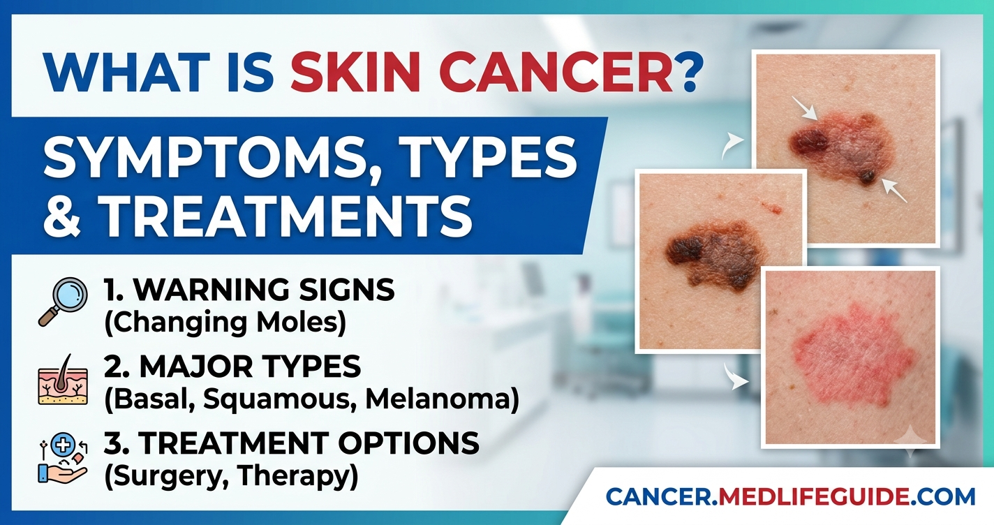
Executive Summary
- Definition & Prevalence: Skin cancer is the out-of-control growth of abnormal skin cells usually triggered by ultraviolet (UV) radiation from sunlight or tanning beds. It is the most common form of cancer in the United States.
- Three Major Types: The most common types are basal cell carcinoma (BCC) and squamous cell carcinoma (SCC) which rarely spread. Melanoma is less common but much more dangerous because it spreads quickly if not caught early.
- Early Detection Saves Lives: Almost all skin cancers are highly treatable when detected early through regular skin self-exams and routine dermatologic evaluations.
EMERGENCY WARNING: If you notice a dark spot on your skin that is rapidly growing bleeding continuously oozing changing colors dramatically or accompanied by swollen lymph nodes persistent fever or shortness of breath seek immediate medical evaluation. Melanoma and advanced skin cancers can spread quickly to internal organs.
What Is Skin Cancer?
Skin cancer is a disease in which skin cells grow abnormally and out of control. Under healthy conditions your skin produces new cells to replace old damaged ones in a orderly process. When skin cell DNA suffers damage—most commonly from ultraviolet (UV) light—the normal cell cycle breaks down. Damaged cells begin to multiply rapidly without dying forming tissue masses known as tumors.
Your skin is your body’s largest organ. It protects you against infection regulates body temperature stores water and fat and manufactures vitamin D. The skin has three main layers:
- Epidermis: The outermost layer of skin that you can see and touch.
- Dermis: The middle layer containing blood vessels nerves hair follicles and sweat glands.
- Hypodermis (Subcutaneous tissue): The deepest layer made of fat and collagen.
Skin cancer originates almost exclusively in the epidermis. The epidermis contains three specific cell types where different cancers form:
- Squamous cells: Flat cells making up the outer surface of the epidermis.
- Basal cells: Round cells located just beneath the squamous cells.
- Melanocytes: Specialized cells located in the deepest part of the epidermis that produce melanin the pigment that gives skin its natural color.
+-------------------------------------------------------------+
|                     EPIDERMIS (Outer Layer)                 |
|  [Squamous Cells]   --> Squamous Cell Carcinoma (SCC)       |
|  [Basal Cells]      --> Basal Cell Carcinoma (BCC)          |
|  [Melanocytes]      --> Melanoma                            |
+-------------------------------------------------------------+
|                     DERMIS (Middle Layer)                   |
|  Blood vessels nerves hair follicles sweat glands        |
+-------------------------------------------------------------+
|                     HYPODERMIS (Deep Layer)                 |
|  Fat connective tissue vascular networks                  |
+-------------------------------------------------------------+
How Skin Cancer Develops: The Role of UV Radiation
The primary cause of skin cell damage is ultraviolet (UV) radiation. UV light is an invisible form of energy produced by the sun and by artificial sources such as indoor tanning beds.
UV radiation is divided into three types:
- UVA rays: These rays penetrate deep into the dermis layer. They cause premature skin aging wrinkles and long-term indirect DNA damage.
- UVB rays: These rays have higher energy levels and directly attack the DNA in epidermal skin cells. UVB rays are the main cause of sunburns.
- UVC rays: These rays are blocked by Earth’s atmosphere and do not reach the ground.
When UV rays strike your skin they can break the chemical bonds inside cellular DNA. Over time repeated sun exposure or blistering sunburns overload the skin’s natural DNA repair mechanisms. Mutations accumulate in critical genes that control cell growth. Once these control genes malfunction damaged cells begin to divide without stopping resulting in skin cancer.
The Main Types of Skin Cancer
Skin cancers are divided into two main categories: non-melanoma skin cancers and melanoma. Non-melanoma skin cancers make up over 95% of all diagnosed cases.
                         Skin Cancer Types
                                 |
       +-------------------------+-------------------------+
       |                                                   |
Non-Melanoma Skin Cancers                              Melanoma
       |                                                   |
       +-------------------+-------------------+           |
       |                   |                   |           |
Basal Cell Carcinoma   Squamous Cell      Rare Types     Melanoma
    (BCC ~80%)       Carcinoma (SCC ~20%) (Merkel etc.) (Most Serious)
Basal Cell Carcinoma (BCC)
Basal cell carcinoma is the most common form of skin cancer accounting for roughly 80% of all diagnoses. It starts in the basal cells at the base of the epidermis.
- Where it appears: Usually on sun-exposed areas like the face head neck shoulders and arms.
- What it looks like: A shiny pearly or waxy bump; a flat firm flesh-colored patch; or a pink sore that bleeds easily and crusts over repeatedly.
- Behavior: BCC grows slowly and rarely spreads (metastasizes) to other parts of the body. However if left untreated it can grow deep into nearby tissue muscle and bone causing severe local damage.
Squamous Cell Carcinoma (SCC)
Squamous cell carcinoma is the second most common skin cancer accounting for about 20% of cases. It forms in the flat squamous cells near the skin’s surface.
- Where it appears: Sun-exposed regions including the ears lower lip face scalp neck hands and lower legs.
- What it looks like: A firm red nodule; a rough scaly flat patch that may bleed; or an open sore that does not heal.
- Behavior: SCC grows faster than BCC and has a small risk (2% to 5%) of spreading to regional lymph nodes and internal organs if ignored.
Melanoma
Melanoma is the most dangerous type of skin cancer. Although it accounts for less than 5% of all skin cancer cases it causes the vast majority of skin cancer deaths. It develops in melanocytes—the pigment-producing cells.
- Where it appears: Anywhere on the body including areas that never see sunlight such as the soles of the feet palms of the hands under fingernails and genital regions.
- What it looks like: A mole that changes shape size or color or a new dark irregularly shaped spot on the skin.
- Behavior: Melanoma can invade nearby tissues and spread rapidly through the bloodstream or lymphatic system to distant organs like the lungs liver and brain.
Rare Skin Cancers
- Merkel Cell Carcinoma: A rare aggressive skin cancer that appears as a firm painless red or purple nodule on sun-exposed skin.
- Sebaceous Gland Carcinoma: An uncommon cancer originating in the oil-producing glands of the skin most often on the eyelids.
- Kaposi Sarcoma: A rare vascular skin tumor caused by a virus (HHV-8) occurring mainly in people with weakened immune systems.
Early Warning Signs: How to Spot Skin Cancer
Detecting skin cancer early gives you the best chance of successful treatment. You should perform a monthly self-exam to check your skin for new spots or changes in existing spots.
The ABCDE Rule for Melanoma
Dermatologists use the ABCDE acronym to help patients identify potential melanomas:
| Letter | Feature | Warning Sign |
|---|---|---|
| A | Asymmetry | One half of the spot or mole does not match the visual appearance of the other half. |
| B | Border | The edges are irregular ragged notched blurred or poorly defined. |
| C | Color | The color is uneven with shades of brown black pink red white or blue. |
| D | Diameter | The spot is larger than 6 millimeters across (about the size of a pencil eraser) though melanomas can sometimes be smaller. |
| E | Evolving | The spot is changing in size shape color or height or develops new symptoms like itching bleeding or crusting. |
                       The ABCDEs of Melanoma

  Asymmetry       Border           Color           Diameter        Evolving
  (Uneven)        (Jagged)        (Mixed)         (>6mm)           (Changing)
    ____            ____            ____            ____            ____
   /    \          /~~~~\          /@ o \          / 6mm\          / ->  \
  |  o   |        |      |        | o  @|         |      |        |  (o)  |
   \____/          \____/          \____/          \____/          \____/
Other Non-Melanoma Warning Signs
- Non-Healing Sores: A sore that bleeds oozes or crusts over stays open for more than 4 weeks or heals and then bleeds again.
- Persistent Pink Patches: A rough scaly patch with a raised border that may itch or hurt.
- Pearly Bumps: A translucent smooth bump that shows tiny blood vessels on its surface.
Unique Clinical Takeaways
To make informed decisions about skin health patients and caregivers need deeper clinical insights beyond standard symptom checklists:
Acral Lentiginous Melanoma (ALM) in Darker Skin Tones
A widespread misconception is that individuals with dark skin tones (Fitzpatrick Skin Types IV–VI) are immune to skin cancer. While their overall risk of UV-induced skin cancer is lower due to higher baseline melanin when melanoma occurs in dark-skinned individuals it is frequently Acral Lentiginous Melanoma (ALM). ALM is not related to sun exposure and develops on non-sun-exposed areas such as the palms of the hands soles of the feet and underneath fingernails or toenails (appearing as a dark longitudinal stripe). Because ALM is often mistaken for a bruise wart or fungal infection it is frequently diagnosed at advanced stages resulting in lower overall survival rates.
The “Ugly Duckling” Concept
While the ABCDE criteria are essential clinical research highlights the Ugly Duckling Sign as a powerful tool for catching unusual melanomas. The core clinical concept is that a patient’s normal moles tend to resemble one another in pattern shade and structure. An abnormal mole or lesion that stands out as completely different from all surrounding moles—the “ugly duckling”—warrants an immediate professional dermatologic biopsy regardless of whether it meets all standard ABCDE parameters.
Mohs Micrographic Surgery and Margin Control
For skin cancers on cosmetic or tissue-sparing areas like the face nose eyelids and ears Mohs micrographic surgery provides the highest cure rate (up to 99% for primary tumors) while preserving the maximum amount of healthy tissue. Unlike standard surgical excision where a surgeon cuts out the tumor and sends it to a lab for results days later Mohs surgery is performed in stages while the patient waits. The surgeon removes a thin layer of tissue maps it and immediately examines 100% of the surgical margins under a microscope in an on-site lab. If cancer cells remain at any edge the surgeon removes another thin layer only from that exact spot minimizing scarring and ensuring complete cancer removal.
Risk Factors: Who Is Most at Risk?
Anyone of any skin color can develop skin cancer. However specific risk factors increase your overall likelihood of developing the disease:
                          Risk Factors Matrix

  High UV Exposure          Fair Skin Features         Medical History
  ------------------        -------------------        -----------------
  * Sunburn history         * Light skin tone          * Personal history
  * Tanning bed use         * Blue/green eyes          * Family history
  * High-altitude living    * Red/blond hair           * Immunosuppression
  * Outdoor occupations     * Freckling skin           * Actinic keratosis
- Sun Exposure and Tanning: A history of multiple severe blistering sunburns—especially during childhood or adolescence—significantly increases melanoma risk in adulthood. Using indoor tanning beds boosts the risk of melanoma by up to 75% when started before age 35.
- Fair Complexion: People with less natural melanin have less built-in protection against UV radiation. Having blond or red hair light-colored eyes fair skin and a tendency to burn easily increases your risk.
- Family or Personal History: If a first-degree relative (parent sibling child) has had skin cancer your risk is substantially higher. Additionally having had one skin cancer increases your risk of developing a second skin cancer.
- Atypical Moles: Having more than 50 ordinary moles or having dysplastic nevi (unusual large irregularly shaped moles) increases your risk of melanoma.
- Precancerous Lesions: Having actinic keratoses—rough scaly pink-to-brown spots caused by sun damage—indicates an elevated risk of developing squamous cell carcinoma.
- Weakened Immune System: Individuals with compromised immune systems—such as organ transplant recipients people living with HIV or patients taking immunosuppressive medications—are at significantly higher risk of aggressive skin cancers.
How Doctors Diagnose Skin Cancer
If you or your primary doctor discover a suspicious spot you will be referred to a dermatologist for formal evaluation.
Visual Dermoscopy
The dermatologist examines the lesion using a dermatoscope—a specialized handheld magnifying device equipped with polarized lighting. Dermoscopy allows the doctor to view sub-surface skin structures pigment patterns and blood vessels that are invisible to the naked eye.
Skin Biopsy
If the doctor suspects cancer they will perform a skin biopsy. The area is numbed with a local anesthetic so you will not feel pain. The doctor removes part or all of the suspicious growth. Common biopsy methods include:
- Shave Biopsy: The doctor shaves off the top layers of the lesion using a small blade. Used mainly for superficial growths.
- Punch Biopsy: The doctor uses a small circular tool to remove a deeper cylinder of skin including the dermis.
- Excisional Biopsy: The doctor uses a scalpel to remove the entire tumor along with a safety margin of normal skin. This is the preferred method for suspected melanomas.
                     Skin Biopsy Procedures

   Shave Biopsy            Punch Biopsy          Excisional Biopsy
   (Surface Layer)        (Dermis Cylinder)        (Full Removal)
       ______                  ______                  ______
      /======\                |  ||  |                /  ||  \
     |________|               |  ||  |               |___||___|
The removed tissue sample is sent to a pathology lab where a pathologist examines the cells under a microscope to provide a definitive diagnosis.
Staging and Further Testing
For basal cell carcinoma and early squamous cell carcinoma staging tests are rarely needed. However for melanoma or high-risk squamous cell carcinoma your doctor may perform staging to see if the cancer has spread:
- Sentinel Lymph Node Biopsy (SLNB): A procedure to check if cancer cells have reached the nearest lymph nodes.
- Imaging Scans: CT scans PET scans or MRI scans to check internal organs for cancer spread.
Staging ranges from Stage 0 (cancer is confined to the top layer of skin) to Stage IV (cancer has spread to distant organs).
Treatment Options for Skin Cancer
Your care team will tailor your treatment based on the cancer type size depth location and stage as well as your overall health.
                       Treatment Modalities
                                 |
       +-------------------------+-------------------------+
       |                                                   |
Local Treatments                                Systemic Treatments
  * Surgical Excision                             * Immunotherapy
  * Mohs Surgery                                  * Targeted Therapy
  * Cryotherapy / Curettage                       * Chemotherapy
  * Topical Creams / PDT                          * Radiation Therapy
Surgical Treatments
- Surgical Excision: The surgeon cuts out the tumor along with a margin of healthy surrounding tissue to ensure no cancer cells are left behind.
- Mohs Micrographic Surgery: Layer-by-layer removal with immediate microscopic examination. Ideal for facial cancers large tumors or recurring cancers.
- Curettage and Electrodessication: The surgeon scrapes away the main tumor using a cup-shaped tool (curet) then uses an electric needle to cauterize the base killing remaining cancer cells and stopping bleeding.
Non-Surgical Local Treatments
- Cryotherapy: Liquid nitrogen is sprayed onto the growth to freeze and destroy cancer cells. Commonly used for actinic keratoses and very small superficial skin cancers.
- Topical Medications: Prescription creams (such as fluorouracil [5-FU] or imiquimod) applied directly to the skin to treat superficial basal cell carcinomas or actinic keratoses.
- Photodynamic Therapy (PDT): A light-sensitizing medication is applied to the skin followed hours later by exposure to a special light source that activates the drug to destroy cancer cells.
Advanced and Systemic Treatments
For advanced recurrent or metastatic skin cancer (especially Stage III/IV melanoma) systemic therapies are used:
- Immunotherapy: Medications (such as immune checkpoint inhibitors) that boost your body’s immune system to recognize and attack cancer cells.
- Targeted Therapy: Drugs designed to target specific genetic mutations in cancer cells (such as BRAF gene mutations in melanoma) stopping their growth.
- Radiation Therapy: High-energy rays used to kill cancer cells in areas where surgery is difficult or after surgery to lower the chance of recurrence.
- Chemotherapy: Strong medications taken orally or intravenously to destroy fast-growing cancer cells.
Prevention and Daily Sun Safety
Up to 90% of non-melanoma skin cancers and 86% of melanomas are preventable through proper sun protection.
Practice the Sun-Smart Rules
- Seek Shade: Avoid direct sun exposure between 10:00 AM and 4:00 PM when UV rays are strongest.
- Wear Broad-Spectrum Sunscreen: Apply a broad-spectrum water-resistant sunscreen with an SPF of 30 or higher every day even on cloudy days. Reapply every 2 hours or immediately after swimming or sweating.
- Wear Protective Clothing: Put on tightly woven long-sleeved shirts and long pants. Wear a wide-brimmed hat that shades your face ears and neck and wear UV-blocking sunglasses to protect your eyes.
- Avoid Tanning Beds: Completely avoid artificial UV exposure from tanning beds booths and sunlamps.
                     Daily Protection Checklist

  [X] Broad-Spectrum Sunscreen (SPF 30+)
  [X] Wide-Brimmed Hat & Sunglasses
  [X] UV-Protective Clothing
  [X] Seek Shade (10 AM - 4 PM)
  [X] Avoid Tanning Beds Entirely
Frequently Asked Questions (FAQs)
Can skin cancer spread to other parts of the body?
Yes skin cancer can spread if left untreated. While basal cell carcinoma rarely spreads melanoma and advanced squamous cell carcinoma can spread to nearby lymph nodes and internal organs.
Is skin cancer fatal?
Most skin cancers (like basal cell and squamous cell carcinomas) are highly curable when caught early. Melanoma is more serious and can be fatal if diagnosed at an advanced stage making early detection critical.
Does sunscreen completely prevent skin cancer?
Sunscreen significantly reduces your risk of skin cancer by blocking harmful UV radiation but it does not offer 100% protection. You should combine sunscreen with shade protective clothing and hat wear.
How often should I have my skin checked by a doctor?
Adults should perform a monthly self-exam at home and see a dermatologist annually for a professional full-body skin exam especially if they have a personal or family history of skin cancer.
Can people with dark skin tones get skin cancer?
Yes people of all skin tones can get skin cancer. While individuals with dark skin have lower overall rates of sun-related skin cancer they are more susceptible to Acral Lentiginous Melanoma on the palms soles and nail beds.